
Ciliate under microscope YouTube
Lab #3 Ciliates under Compound Microscope. 9/7/17. Purpose: The purpose of this lab is to allow students to observe ciliates in greater detail with the higher magnification of the compound microscopes, compared to the dissecting microscopes. students will also get the opportunity to practice operating a compound microscope. Materials:

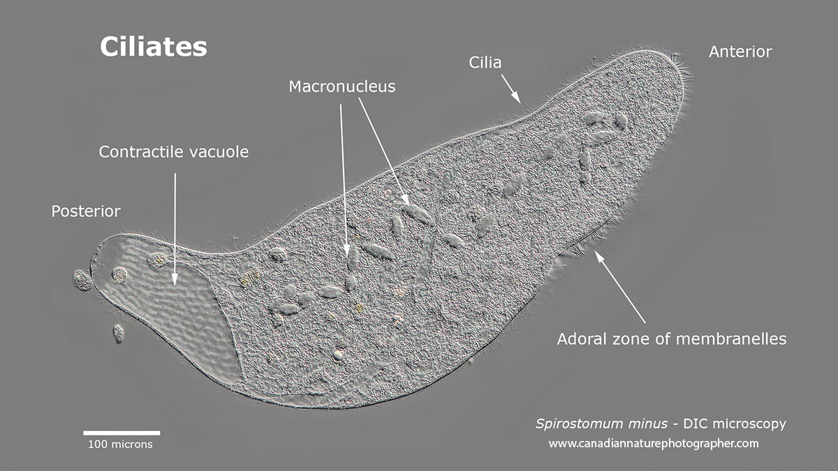
Photographing Ciliates The Canadian Nature Photographer
Place a drop of pond water under the microscope, and you will likely find an ocean of extraordinary and diverse single-celled organisms called ciliates. This remarkable group of single-celled organisms wield microtubules, active systems, electrical signaling, and chemical sensors to build intricate geometrical structures and perform complex behaviors that can appear indistinguishable from.
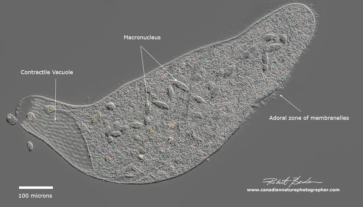
Photographing Ciliates The Canadian Nature Photographer
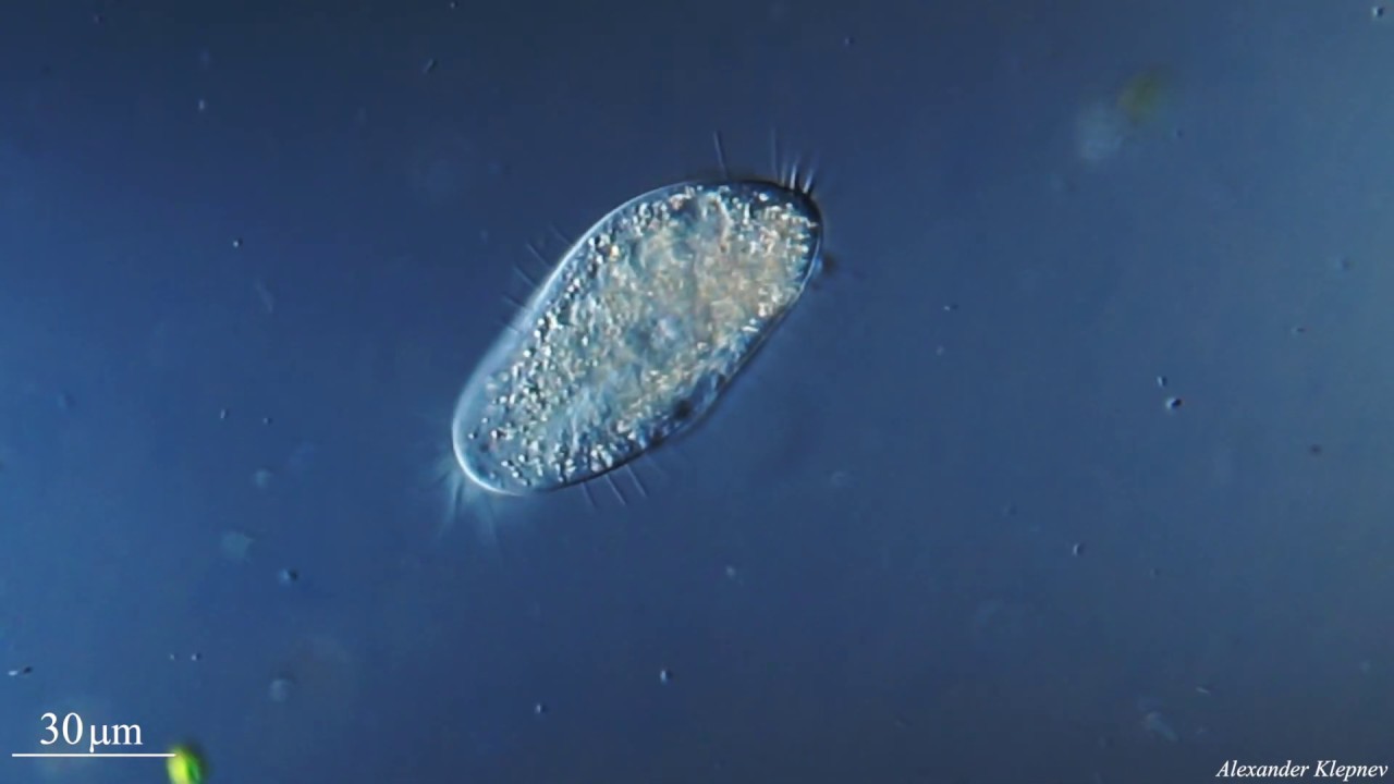
The ciliates are so named because of the cilia, small hairs that are distributed over the entire body. Ciliates are generally ovoid or pear-shaped and maintain their shape by means of a tough but flexible pellicle. Cilia protrude through the pellicle in a variety of patterns.. When counting flagellates under the microscope, a 400X.

Ciliate under microscope YouTube
This special issue of the Journal of Eukaryotic Microbiology (JEM) summarizes achievements obtained by generations of researchers with ciliates in widely different disciplines. In fact, ciliates range among the first cells seen under the microscope centuries ago. Their beauty made them an object of.
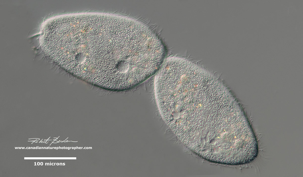
Photographing Ciliates The Canadian Nature Photographer
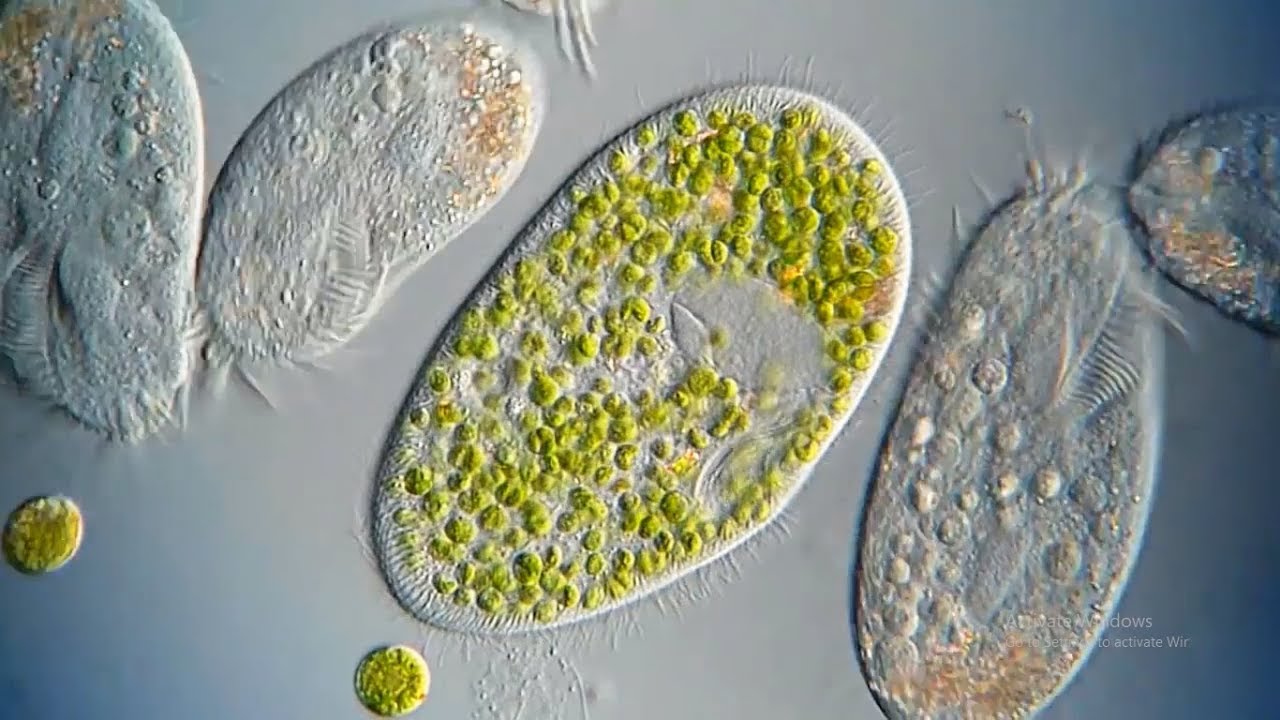
Habitats. Ciliates are divided into free living and parasitic. Whereas free living ciliates (can live outside a host) can be found in just about any given environment, parasitic ciliates live in the body of the host. Paramecium is an example of free living. Such paramecia as Paramecium caudatum can be found free living in fresh water bodies.
The Micro Universe Microscopic life by Robert Berdan The Canadian
The ciliates are a group of alveolates characterized by the presence of hair-like organelles called cilia, which are identical in structure to eukaryotic flagella, but are in general shorter and present in much larger numbers, with a different undulating pattern than flagella. Cilia occur in all members of the group (although the peculiar Suctoria only have them for part of their life cycle.

Ciliates under the microscope YouTube
ciliate, any member of the protozoan phylum Ciliophora, of which there are some 8,000 species; ciliates are generally considered the most evolved and complex of protozoans. Ciliates are single-celled organisms that, at some stage in their life cycle, possess cilia, short hairlike organelles used for locomotion and food gathering.. The cilia are usually arranged in rows, known as kineties, on.

Ciliates Under Microscope
How ciliates got their nuclei. Biologists who spend time observing environmental samples under the microscope are used to the incredible range of shapes, sizes, and behaviors displayed by eukaryotic microorganisms, which rivals or exceeds that of animals, just on a smaller scale. These are lumped together as "protists" and generally.

Phylogeny of the ciliate family Psilotrichidae (Protista, Ciliophora
Place this slide under the microscope and observe clearer images and descriptions of the ciliate you are able to gather using the different magnifications of the microscope. Repeat this process as many times as necessary to gain accurate observations and pictures of the ciliates. Record all of your observations.

4K Blepharisma ciliate under the microscope YouTube
Live ciliates were observed for morphological details using differential interference contrast (DIC) microscopy with a Leitz (Weitzlar, Germany) microscope at a magnification of 300-1250 × with the help of a compression device . For examination of the swimming behavior, ciliates were observed in a glass depression slide (3 ml) under a.

Ciliates Under Microscope
Ciliates are basically ciliated protozoans. Protozoans are another term for a group of single-celled eukaryotes. They are either parasitic or free-living and feed on organic matter such as debris, organic tissues, or other microorganisms. Contents show.
Under the microscope a ciliate YouTube
Ciliates are recovering from a piece of frozen, dried mosses and wandering around.
Ciliate under microscope YouTube
The Litostomatea are a class of ciliates. The group consists of three subclasses: Haptoria, Trichostomatia and Rhynchostomatia. Haptoria includes mostly carn.
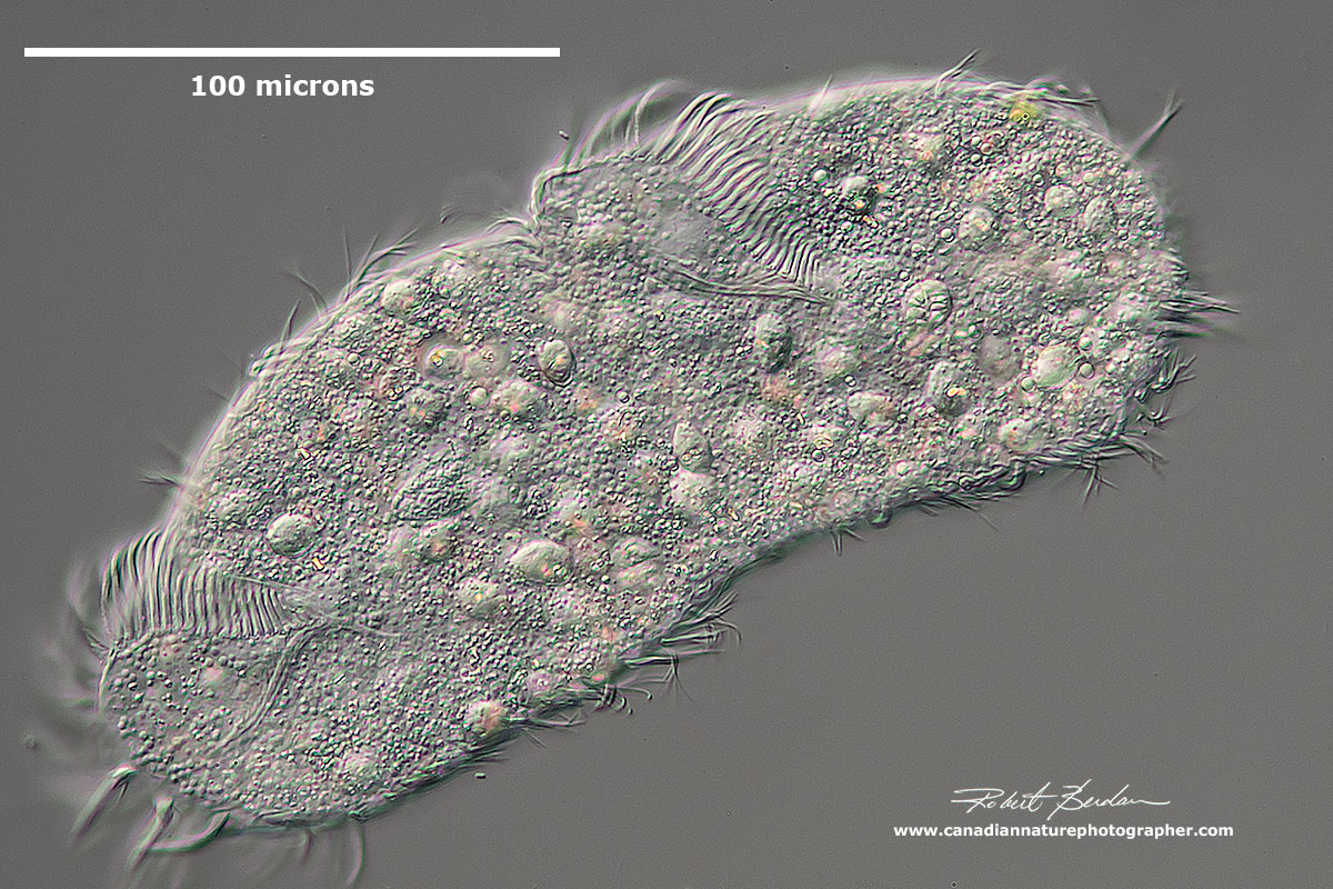
Photographing Ciliates The Canadian Nature Photographer
[In this video] A video collection of several common ciliates under the microscope. Larger eukaryotes, such as animals, have cilia as well. Cilia are usually present on a cell's surface in large numbers and beat in coordinated waves. In humans, cilia are found on the epithelial cells lining the respiratory tract. These cilia move constantly.
What are Ciliates? YouTube
This video shows a species of Opercularia. These are ciliates that are found attached to surfaces (they are sessile), and that typically live in colonies. He.

ciliates 400x magnification YouTube
This video shows a ciliate; a type of single-celled organism that inhabits a wide range of freshwater habitats. Ciliates feed upon smaller microscopic organi.
