
C3C4 Posterior Cervical Laminectomies, C67 Laminotomy and Spin Stock
A 59-year-old man underwent four-level (C3-C4, C4-C5, C5-C6, C6-C7) discectomy and fusion with a PEEK cage filled with autogenous iliac cancellous graft due to cervical spondylotic myelopathy. Two-year postoperative follow-up lateral X-ray showing complete bony fusion and maintenance of the cervical lordotic curve.
Spine Surgery Cervical Disc Protrusions at C45 and C56 with Stock
A pinched nerve or nerve root inflammation may also cause numbness and/or weakness to radiate down into the shoulder, arm, hand, and/or fingers. Radicular pain may also accompany radiculopathy in some instances. See Cervical Radiculopathy from a Herniated Cervical Disc Symptoms worsen with specific head positions or activities.
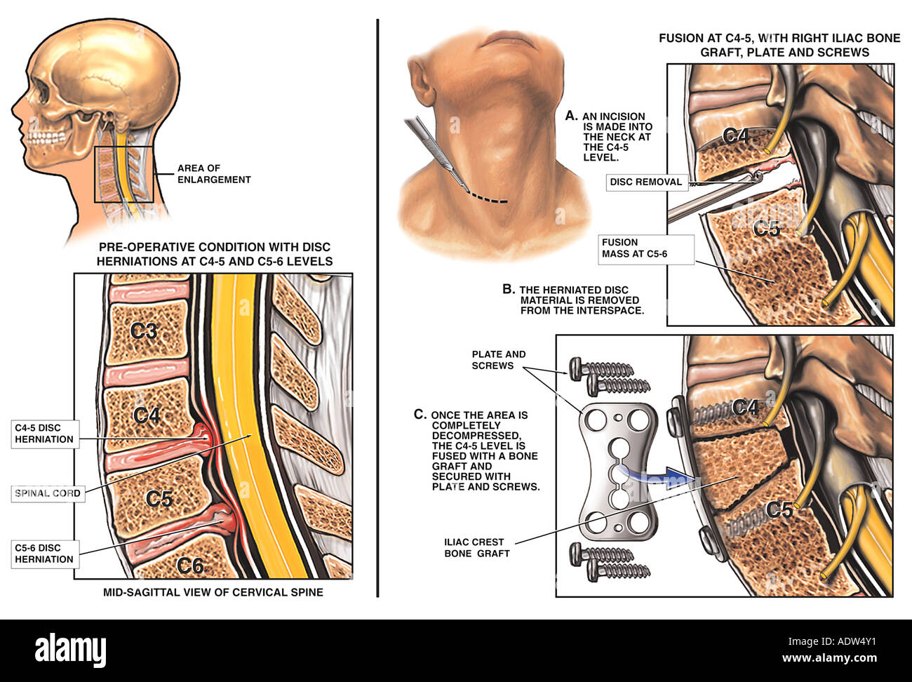
C45 and C56 Anterior Cervical Discectomy and Fusion Stock Photo
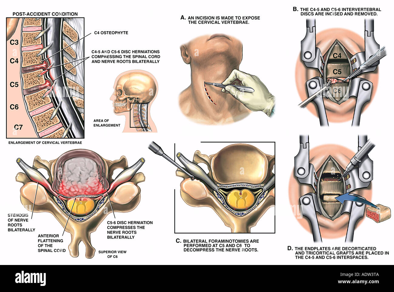
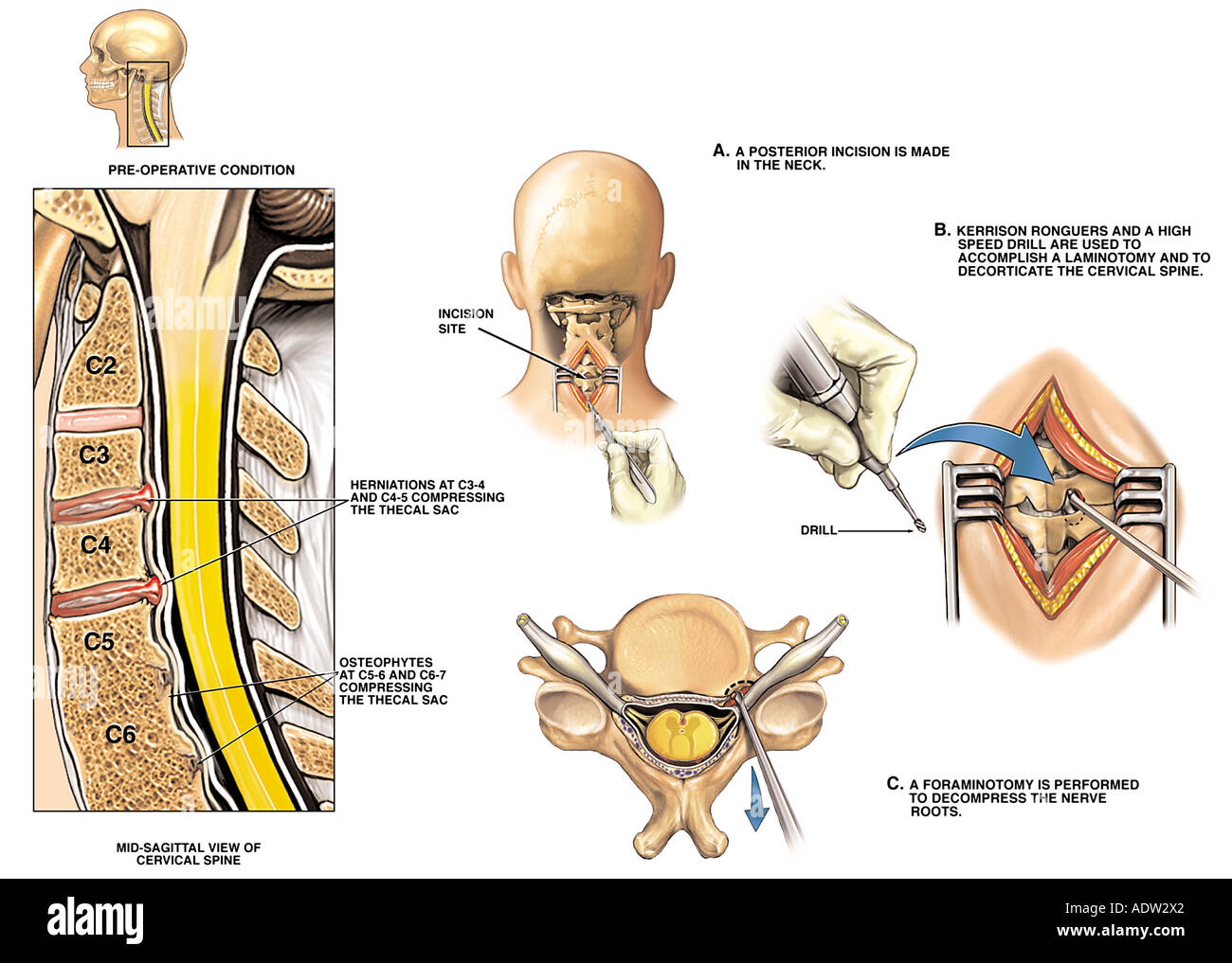
The operation proceeded with the patient in a prone position with midline dissection to the posterior elements of C3-C6. Pilot holes were drilled in the lateral masses and C3-C4, C4-C5, C5-C6, and C6-C7 laminectomies performed using a high-speed drill, with removal of the laminae en bloc. Lateral mass screws were inserted and precontoured rods.

La chirurgie de la colonne vertébrale C4C5 et C5C6 et du col de l
Cervical Annular Tears (C1-C7) Thoracic Annular Tears (T1-T12) Lumbar Annular Tears (L1-L5) Types of Annular Tears. Peripheral Tears - An injury such as a fall or car accident is the most common cause of this type of annular tear. These tears begin on the outside layer of the discs that surround the disc.
Puesto condición de accidente con espondilosis Osteophytes y la
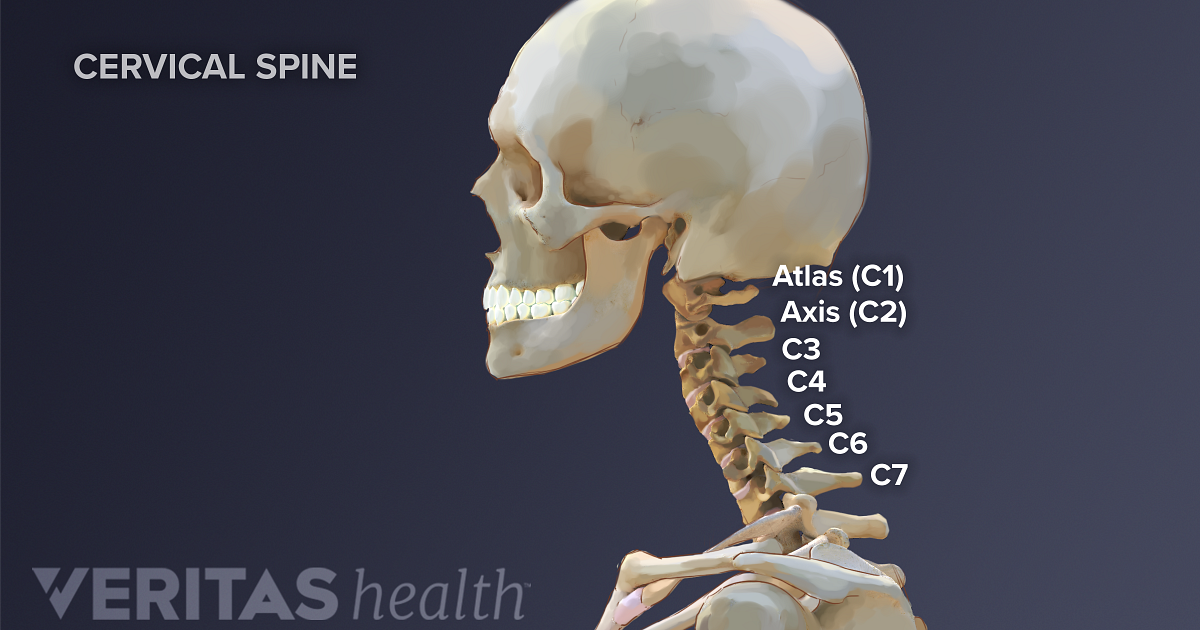
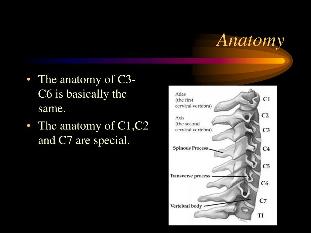
Your seven cervical vertebrae (C1 to C7) are connected at the back of the bone by a type of joint (called facet joints), which allow for the forward, backward and twisting motions of your neck. Your cervical spine is also surrounded by muscles, nerves, tendons and ligaments.

Neck Surgery C5 C6 C7 Recovery Time Renew Physical Therapy
Cervical discs have three purposes: cushioning the force applied to the spine to protect the vertebrae from damaging each other. acting as connective tissue to keep the cervical discs in place. acting as a cartilaginous joint to allow us to have a range of motion between bones to turn or bend the spine.
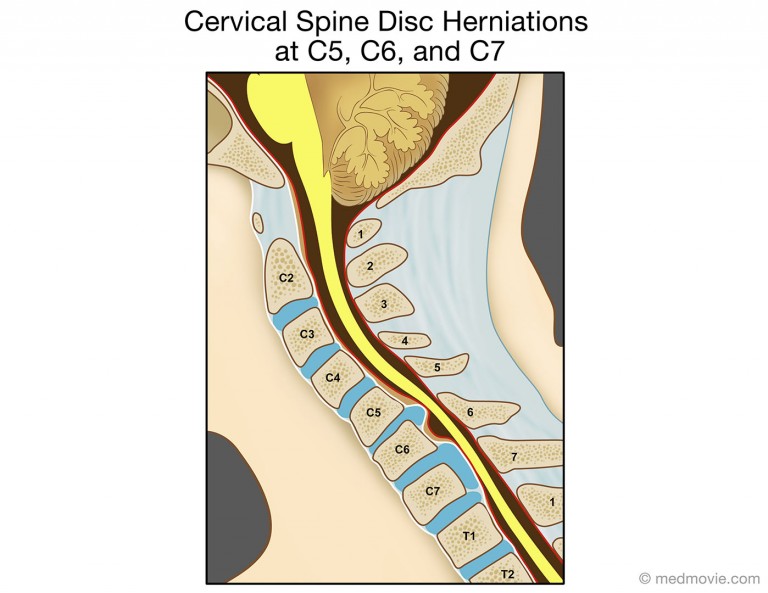
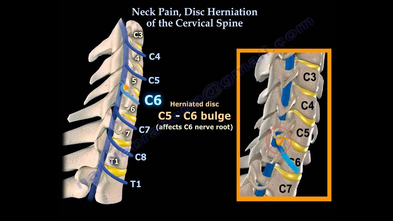
Cervical Spine Disc Herniations at C5, C6, and C7
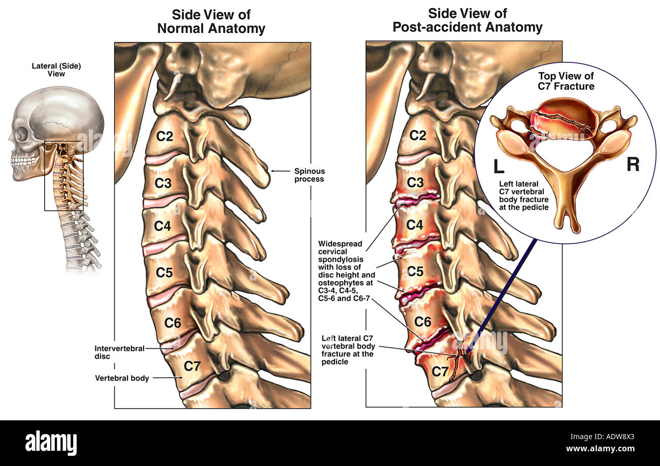
Anterolisthesis of C3 on C4 and C4 on C5. Vertebral body heights are maintained. Multilevel degenerative changes characterized by disc space loss, uncovertebral joint degeneration, and facet arthropathy, most pronounced at C6-C7. The prevertebral soft tissues are within normal limits.

Sagittal MRI image showing C4C5,C5C6,C6C7 disc herni Openi
If pain and symptoms from cervical degenerative disc disease intensify, steps can be taken to help alleviate the problem. The first steps are usually self-care and/or non-surgical treatment options, and typically these will effectively manage the pain.
slip disc cervical spondylosis Max Short
There is a severe narrowing of the diameter of the spinal canal at C4-C5 and less prominent at C5-C6. Getty Images What Causes Cervical Spinal Stenosis? A common cause of cervical spinal.
Neck Injuries C34, C45 Disc Herniations and C57 Osteophytes Stock
Cervical osteophytes are bone spurs that grow on any of the seven vertebrae in the cervical spine (neck), ranging from the base of the skull, C1 vertebra, to the base of the neck, C7 vertebra. Show Transcript The term "bone spurs" might elicit images of radiating spikes, but bone spurs (osteophytes) are actually rounded and scalloped.
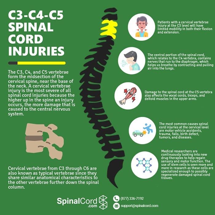
C3, C4, & C5 Vertebrae Spinal Cord Injury
Cervical Spine The neck is part of a long flexible column, known as the spinal column or backbone, which extends through most of the body. The cervical spine (neck region) consists of seven bones ( C1-C7 vertebrae ), which are separated from one another by intervertebral discs.
All About the C2C5 Spinal Motion Segments
The herniated disk was located at C3-C4, C4-C5 in 1 case, C4-C5, C5-C6 in 11cases, C5-C6, C6-C7 in 7 cases, C4-C5, C6-C7 in 3 cases, and C4-C5, C5-C6, C6-C7 in 4 cases. There were 12 cases with myelopathy and 14 patients with radiculopathy. The stabilization and the range of motion of implanted disk, the fusion of cage, and the displacement of.
PPT Practical approach to Cervical Spine Trauma PowerPoint
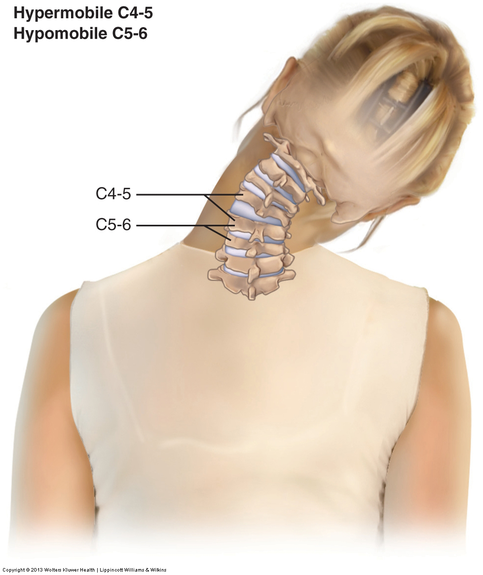
The degenerative disc disease of the cervical spine usually involves the most mobile segment that is the C5-C6 followed by C6-C7 and C4-C5 disc levels. The degeneration causes decreased water content of the disc or desiccation which leads to tears in the outer ring or the annulus fibrosus.
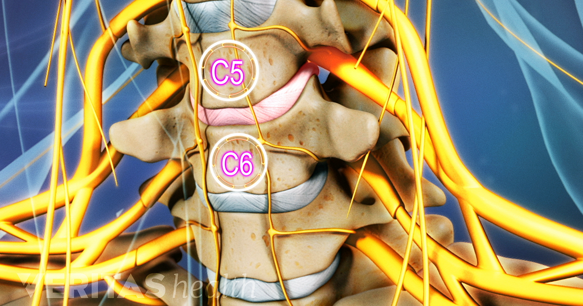
All About the C5C6 Spinal Segment
Degenerative disc disease of the cervical spine typically develops in the aging population equally in terms of patient sex. Patients most commonly present with pain. Pain, or in combination with other neurological symptoms, may require surgical intervention.

Cervical Vertebrae Vector Illustration Scheme With Skull C1 Atlas C2
Overview Cervical spondylosis is a general term for age-related wear and tear affecting the spinal disks in your neck. As the disks dehydrate and shrink, signs of osteoarthritis develop, including bony projections along the edges of bones (bone spurs). Cervical spondylosis is very common and worsens with age.
Joint Dysfunction (subluxation, misalignment) of the Cervical Spine
C3, C4, C5, and C6 cervical vertebrae share characteristics with most of the vertebrae throughout the spine. Cervical vertebrae C3 through C6 are known as typical vertebrae because they share the same basic characteristics with most of the vertebrae throughout the rest of the spine. Typical vertebrae have: Vertebral body.